You must be signed in to read the rest of this article.
Registration on CDEWorld is free. You may also login to CDEWorld with your DentalAegis.com account.
The economics of the baby boom—exhaustively studied and thoroughly parsed—offer lessons for problems facing dentistry today. Growth fueled by the group born between 1946 and 1964 proved a boon to dental services across a wide breadth, including boutique or spa care, implants, cosmetic innovations, hygienist-dispensed periodontal products, and comprehensive oral rehabilitation. In addition, advances in pain management and sedation have compelled adults to embrace long-postponed procedures.
Now the dental profession faces another defining moment, carrying similarly deep potential. The current challenge is caused by an explosion in the number of children in the United States—totaling 24% of the population, numbering approximately 74 million.1 The needs of this population are both complementary and unique when contrasted with their parents, and dentists should prepare and adapt accordingly.
The Caries Explosion
Some aspects of the present environment are surprising. After decades of advances in preventive technology, dental disease remains unconquered. The caries rate is rising in preschool children, especially among those from lower and middle socioeconomic backgrounds. According to recent studies by the U.S. Centers for Disease Control and Prevention (CDC), more than 4 million preschoolers are affected by dental caries, a jump of 600,000 in 10 years.2 For children ages 2 to 5, the caries rate was 24% between 1988 and 1994. The number rose to 28% between 1999 and 2004.3,4 Will this trend continue, and will these same children incur even more caries as they grow older? It is not over-reaching to anticipate other consequences. Today an estimated 80% of all American adolescents have tooth decay by the age of 17.5 .
Many counties in the United States cite tooth caries as the single largest unmet health issue, even placing it ahead of asthma, diabetes, obesity, and heart problems. An estimated 51 million school hours are lost each year due to poor oral health. Dental-related emergencies often constitute the first “dental visit” for 25% of children seen in a children’s hospital.6
Unfortunately, a dwindling base of dental practitioners is accompanying this skyrocketing demand for pediatric care. There are approximately 183,000 active licensed dentists in the country, and 80% are general practitioners. Only 4%—about 8,000—are pediatric dentists. Postdoctoral programs in pediatric dentistry graduate a little over 300 new specialists per year, with some returning to their own countries.7
Given this disturbing disparity, general practitioners must play a major role along with the pediatric dentist in delivering care to children. Clearly the two must partner in this response. The question is not whether general dentists should deliver pediatric services but rather how they can be trained to do so selectively and well.
Complicating Factors in Pediatric Dentistry
The increase of caries in children combined with the stresses of harried and time-strapped parents adds to the complexity of treating this population. However, just as sedation dentistry enhances the comfort level of adults and permits longer appointment times that allow more procedures, so can children receive these benefits. Regardless of patient age, though, careful case selection and proper training are critical prerequisites to the delivery of safe sedation dentistry.
Also akin to adult care, sedation is a natural extension of compassionate treatment because it offers palliative or anxiety-reducing measures. Yet pain control for children has historically been both underreported and poorly addressed. Until relatively recently, children too young to speak were also believed to be too young to experience pain or fear—and as a result they were not likely to be administered analgesia, even after major surgery. Among the reasons for this was misunderstanding about safe pediatric medication and appropriate dosage.8
As for the spectrum of pediatric sedation, it runs the gamut, including minimal, moderate, and deep levels along with general anesthesia. These categories differ through their effects upon the central nervous system, not the route of administration.
A number of administration routes are employed in pediatric sedation. In enteral administration, a sedative agent is absorbed through the gastrointestinal tract or oral mucosa (oral, sublingual, or rectal). The sedative agent bypasses gastrointestinal absorption in parenteral administration (intramuscular, intravenous, submucosal, subcutaneous, intranasal, and intraosseous). Transdermal administration involves absorption of the sedative agent through the skin (patch or iontophoresis). In transmucosal administration the sedative agent is absorbed through the mucosa (intranasal, sublingual, or rectal). The sedative agent is absorbed through the lungs in inhalation administration because the patient breathes a gaseous form of the agent.
Enteral Sedation
For the purposes of this article, discussion will be limited to enteral—administered orally—minimal sedation. Pediatric minimal sedation is defined by the American Dental Association in its Guidelines for the Use of Sedation and General Anesthesia by Dentists (2007).9 For children under 12, the ADA supports the American Academy of Pediatrics/American Academy of Pediatric Dentists Guidelines for Monitoring and Management of Pediatric Patients During and after Sedation for Diagnostic and Therapeutic Procedures (2006).10
Both organizations characterize pediatric enteral minimal sedation as a minimally depressed level of consciousness, produced by an orally administered pharmacological agent or agents. The child is allowed to independently and continuously maintain the airway, and to respond appropriately and normally to tactile stimulation and verbal commands.
The drug or drugs used along with sedation techniques should afford the child the widest margin of safety; that is, the medication should never render loss of consciousness. Sedatives should only be administered to children in the dental setting, and never be provided for delivery at home prior to the appointment. This prevents the possibility of a compromised airway and respiratory depression during transport.
Even where the intent is to minimally sedate, moderate sedation may still result. In this case the dentist should be prepared to quickly apply guidelines for moderate sedation. Nitrous oxide/oxygen analgesia may be used in conjunction with pediatric oral conscious minimal sedation, but this combination can potentially produce deeper levels of sedation. Local anesthesia can and should be used in conjunction with orally administered sedative drug(s) and nitrous oxide/oxygen analgesia. But this must be used in accordance with the maximum recommended therapeutic dose (MRTD) of the local anesthetic appropriate to the weight and age of the child.
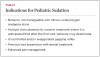
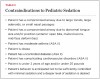
Sedation of a child is a continuum. Thus, it is not always possible to predict how a child will respond. Dentists intending to provide minimal sedation should be able to diagnose and manage physical consequences in the event the level of sedation becomes deeper than expected. For a list of indications and contraindications to the administration of pediatric sedation, see Table 1 and Table 2.
Newer drugs offer an unprecedented level of safety. Previous surveys11,12 reflect the current use of midazolam (Versed) as the most common pediatric single-sedative drug. Midazolam and hydroxyzine (Vistaril, Atarax) have become the most popular combination of sedative drugs.
Midazolam
Midazolam was first synthesized in 1976 by Fryer and Walser and marketed as an ultra short-acting intravenous benzodiazepam derivative.13 Midazolam was often used in combination with other medications for pre-procedural induction or procedural sedation. Midazolam (Versed) became commercially available in 1998 when Roche Pharmaceutical released it in syrup form. It gained popularity for its rapid onset, brief duration, and short half-life. Midazolam’s ability to calm a severely apprehensive young child provided immediate applicability to pediatric dentistry.14
Midazolam shares the pharmacodynamic properties of other benzodiazepines but also provides a relatively short-acting central nervous system depressant15-18 with anxiolytic, hypnotic, anticonvulsant, muscle relaxant, and anterograde amnesic effects. Midazolam has very little analgesic effect.16 Thus, it is supplemented with nitrous oxide/oxygen analgesia.
Midazolam’s effects on the CNS depend upon dose, route of administration, and concomitant use with other medications.15,18 Midazolam is rapidly metabolized and excreted. Its onset of action takes place within approximately 5 to 15 minutes, and its peak plasma levels occur within 20 to 50 minutes. It has a plasma elimination half-life between 2 to 4 hours.19-24 Midazolam is both water-soluble and highly lipid-soluble at the physiologic pH, which allows for rapid entry into brain tissue and as well as a relatively rapid onset of action.18
The oral bioavailability rate of midazolam is approximately 35% to 40% because of the “first-pass” phenomenon. The first-pass effect is defined as the metabolism of orally administered drugs by gastrointestinal and hepatic enzymes, resulting in the biotransformation, reduction, or even elimination of the substance before reaching the circulation.
The primary effect of midazolam appears to be reversible binding to the gamma aminobutyric acid (GABA), an inhibitory neurotransmitter.25 This in turn binds to the GABA receptors on the neuron membrane in the central nervous system.18,24 The result is depression of the level of consciousness; that is because the CNS neurons are hyperpolarized and rendered less responsive to excitation. The routine dose ranges from 0.3 mg/kg to 0.75 mg/kg with the maximum recommended therapeutic dose (MRTD) of 20 mg16 (15 mg in preschool children). Flumazenil (Anexate, Romazicon), an imidazobenzodiazepine derivative, is the GABA-receptor antagonist used as the reversal agent for midazolam. Recommended pediatric dosages of flumazenil are: 0.01 mg/kg IV over 15 seconds as needed to a maximum of 0.2 mg; four maximum repeated doses at 0.005 mg/kg to 0.01 mg/kg (maximum dose of 0.2 mg) at 1-minute intervals. The maximum total cumulative dose should be 0.05 mg/kg or 1 mg, whichever is lower. In the absence of an intravenous approach, an alternative route of administration is submucosal, under the tongue (sublingual).
The major risk of midazolam in higher doses is hypoventilation. It may induce respiratory depression through marked decrease in the ventilator response to carbon dioxide18 similar to that which follows administration of diazepam. This effect is more dramatic in patients with chronic obstructive lung disease.18 Contraindications for midazolam include but are not limited to: hypersensitivity to benzodiazepam-class drugs, acute narrow-angle glaucoma, shock, hypotension, pregnancy, and head injuries.
The biotransformation of midazolam is mediated by the cytochrome P450 3A4 enzyme system in the gastrointestinal tract mucosa as well as the liver.26 Drugs or other dietary foods that inhibit the activities of cytochrome P450 3A4 such as erythromycin, diltiazem, verapamil, ketoconazole, and itraconazole as well as grapefruit juice will inhibit midazolam clearance and prolong plasma clearance.16,27,28 The elimination of midazolam is primarily via the hepatic metabolic metabolism and excreted in the urine.16
Hydroxyzine
Hydroxyzine was first synthesized in the 1950s as an antihistamine and H1 receptor antagonist. It is available as Vistaril and Atarax in oral formulation. This antihistamine is primarily used for its anti-allergenic and anti-emetic properties. It is a weak analgesic by itself but an opioid potentiator with some anxiolytic properties.29 In addition to its effect on the H1 receptor site, hydroxyzine exhibits subcortical CNS suppression, lassitude, decreased mucosal secretions, euphoria, and mild analgesic effects.16,30
Hydroxyzine is a common and remarkably safe medication for the purpose of pediatric sedation. In addition to its sedative effect, hydroxyzine prevents nausea and vomiting, and has drying effects that come from the anti-allergic properties. These make the medication a good adjunct to the benzodiazepine family.
When administered orally, hydroxyzine is absorbed from the gastrointestinal tract and metabolized in the liver. After oral administration, hydroxyzine has an observable onset of clinical effect within 15 to 30 minutes. The maximum effect takes place approximately 1 to 2 hours after oral administration, with the overall effects dissipating in 3 to 4 hours. The half-life is approximately 6 to 10 hours. Other than drowsiness, hydroxyzine has minimal effect on cardiovascular or respiratory function.31,32 The drug carries minimal abuse, dependency, addiction, and toxicity potential when used in the therapeutic range.26 Side effects of hydroxyzine include dizziness, insomnia, tremors, seizures, and urinary retention.30,31
The routine dose for hydroxyzine is 1 mg/kg to 2 mg/kg up to a maximum recommended therapeutic dose (MRTD) of 50 mg for children and up to 100 mg for adults.28,31 Pediatric dentists have used hydroxyzine as a sedative agent safely for many years.33,34
Combining Midazolam and Hydroxyzine
Midazolam and hydroxyzine alone carry limitations. However, the combination of these drugs provides benefits surpassing those of either agent used alone. While midazolam’s rapid onset is advantageous, its working time is moderate. Hydroxyzine has a slower onset but affords a longer duration of action. The combination of the two potentiates their singular effect, thus allowing for a more efficient treatment start time and longer duration.33,35 The combination has quickly become one of the most popular drug regimens in pediatric sedation.
Monitoring Pediatric Sedation
Pediatric sedation dentistry has benefited from extraordinary developments in monitoring technology. Still, nothing can replace the observational skills of the doctor performing sedation. The basic guidelines of “look, listen, feel” should serve as the primary modes of monitoring by dentists and staff when it comes to assessing the effects of the sedative medications on the patient. These remain crucial registers of the well-being of any child undergoing enteral minimal sedation.
The American Academy of Pediatric Dentistry (AAPD) and the American Academy of Pediatrics (AAP) have adopted specific standards for monitoring sedated pediatric dental patients at the levels of minimal, moderate, and deep sedation. The guidelines specify requirements for personnel, monitoring, and documentation before, during, and after the procedure10 in the case of minimal sedation levels, but with competency at moderate levels.
Components of properly monitoring a moderately sedated patient include:
- Practitioner and support person
- Pulse oximeter to measure oxygen saturation and heart rate
- Precordial stethoscope and/or capnography monitor to record intermittent respiratory rate
- Noninvasive device or a sphygmomanometer to assess blood pressure
- Temperature recording
- Documentation of monitoring values on a time-based record
The child’s skin, lips, and mucosal color should be continually observed for any change. The color of these tissues is a prime indicator of oxygen profusion; they should always be pink. The skin of the forehead, cheeks, neck, and chest are not only additional indicators of oxygen profusion, but suggestive of a possible allergic reaction to localized (latex) or generalized etiology (systemic drug). Skin color often indicates an increase in temperature, sweating, and capillary vasodilatation or the presence of petechiae (broken surface blood vessels).
The eyes should be scrutinized for changes in the level of consciousness. If the eyes are opened but the pupils roll backward toward the scalp, or they show signs of lateral strabismus, then verbal command or physical stimulation may be necessary to assess the level of sedation.
The position of the head, neck, and tongue should continually be observed to ensure proper airway maintenance. The tongue is the most frequent source of airway obstruction. The pharyngeal area should be continually assessed to gauge the existence of accumulations of oral secretions and/or esophageal/stomach fluids. Fluids in the posterior pharyngeal area or larynx are common causes of laryngeal spasms.
Respiration depth, breathing, quality, and rate can be continually assessed by observing the movement of the chest and abdomen. This is determined by sight and also via precordial stethoscope, which offers information about the patient’s heart rate and rhythm.
Pulse Oximetry
Pulse oximetry is the measurement of transmitted light through a translucent site. This measurement determines a patient’s oxygenated hemoglobin saturation status noninvasively using spectrophotometry (instruments that measure different wavelengths and intensity of light). It uses measurements obtained from two wavelengths of light in the red and infrared range emitted through the nail bed of the patient’s finger or a toe (with any decorative polish removed). This measurement can also be achieved on other sites, such as an infant’s forehead or earlobe. When arterial oxyhemoglobin saturation is measured by an arterial blood-gas analyzer, it is referred to as SaO2. When arterial oxyhemoglobin saturation is measured noninvasively by pulse oximetry, it is referred to as SpO2.
This instrument carries several advantages with regard to sedation. For example, it detects developing hypoxemia by measuring decreases in the percentage of hemoglobin oxygen saturation (SaO2). Pulse oximetry machines offer information such as SpO2 (oxyhemoglobin), pulse rate, wave-form displays, and alarms, among other features. The pulse oximeter measures the percentage of hemoglobin saturated with oxygen molecules. The percentage of oxyhemoglobin saturation and the affinity of the oxygen molecule to the hemoglobin are charted mathematically as a nonlinear curve described as the “oxyhemoglobin dissociation curve.”
This curve mathematically equates the percentage of saturation of hemoglobin on the vertical axis to the partial pressure of oxygen in the blood on the horizontal axis. The curve shifts to the right and reflects decreased O2 affinity with increased temperature, increased CO2, acidosis, and exercise.
A precordial stethoscope—also called a Wenger chest piece—attaches via double-sided tape to the skin of the patient, usually near the suprastenal notch. The precordial stethoscope allows the dentist to confirm ventilation, quality of breath sounds (ie, wheezing), the regularity of the heart rate, and the quality of heart tones. It also permits detection of any changes or interferences of the airway patency. In the past, an air tube linked the “bell” to the earpiece. Newer technologies and wireless devices such as Bluetooth eliminate the need for a tube.
A pediatric sedation emergency is inevitably associated with an airway complication.36 The use of a precordial stethoscope allows the dentist to recognize and remedy a situation that compromises the airway quickly before desaturation can occur.
Capnography
Capnography monitors carbon dioxide in the respiratory gases using infrared spectrometry. This machine provides an instantaneous and continuous graphic record of carbon dioxide content in expired gases. It also provides a digital recording of the respiration rate.37 This device directly reflects the elimination of CO2 by the lungs; indirectly, this monitor’s data reflects the production of CO2 by the tissue and the circulatory transport of CO2 to the lungs.
A capnography machine thus assists in the monitoring of the metabolic rate of the patient; the end tidal carbon dioxide represents its concentration prior to the initiation of inspiration.38 Sampling air is vacuumed through a port that is taped either below the patient’s nostrils, next to the mouth, or within the scavenger port of the Porter nitrous oxide mask (side-stream sampling), and delivered into the capnography machine for the detection of the presence of CO2. The cycle of breathing is represented by expiratory carbon dioxide. The drawback of the capnography machine is that crying and/or mouth breathing, clinical events that are not uncommon during pediatric enteral sedation, may activate the alarm without the presence of apnea episodes.39,40
Capnography has proved more effective than clinical judgment alone in the early detection of adverse respiratory events such as hypoventilation. During sedation, capnography provides more useful information (such as the frequency and regularity of ventilation) than the pulse oximeter. Capnography is highly accurate in detecting complete airway obstruction or apnea.41 It is also a very useful indicator of hypoventilation and/or hypercarbia.42
Blood pressure is the force of the blood exerted against the walls of the arteries. It is determined not by the strength of the heart muscles but rather by how much resistance exists in the arteries. Each time the heart heats, it pumps out blood into the arteries. The blood pressure is highest when the heart beats, pumping the blood through the circulatory system, the systolic pressure. When the heart is at rest and between beats, the blood pressure falls, the diastolic pressure. The blood pressure can also change throughout the day, falling to its lowest during sleep and rising to the highest during periods of anxiety or activity. Artifacts affecting blood pressure measurement include a too-small cuff, which can result in a reading that is too high. A cuff that is too large will often provide readings that are too low.
A time-based sedation record documents vital-sign values; this should become part of the patient’s permanent record. This legal document indicates the patient’s name, address, and telephone numbers of the medical home; a pre-sedation health evaluation containing the patient’s current age and weight; a current health history and review of systems; current vital signs of heart rate, blood pressure, respiration rate, temperature, documentation of a physical examination, and an assessment of the patient’s health and physical status, or an ASA classification. The sedation record should also document the delivery of any medication, dosage, and the route and time of administration.
The time-based record documents that a formal identification of the patient and confirmation of signed informed consent was performed, the patient medication and dosages, route of administration, the concentration of oxygen and inhalation analgesia being used in conjunction to the oral sedation medication, the duration of administration of inhalation analgesia, level of consciousness at the time of administration and responsiveness, heart rate, blood pressure, respiration rate, oxygen saturation, and any adverse reactions.
The time-based sedation record should note the patient’s level of consciousness after the sedation procedures are terminated. The oxygen saturation in room air should be recorded and should have returned to within 10% of the pre-sedation level. Documentation should also include the patient’s ability to ambulate satisfactorily; to remain upright; to appropriately respond to verbal stimuli, and to recognize themselves and their environment. All monitoring should be continued until the patient has attained predetermined discharge criteria.
Emergency Preparedness
As noted earlier, this article discusses enteral minimal conscious sedation. However, sedation’s effects are unique to each individual and, additionally, revealed along a continuum. Both the practitioner and staff must be prepared to respond when sedation progresses to a higher level than planned or in the event that complications arise. The authors advise (and some states require) that practitioners performing pediatric sedation have emergency preparedness protocols in place encompassing both the sedation team and the physical office. It is also recommended that practitioners be certified in Pediatric Advanced Life Support (PALS) and obtain recertification at least once every 2 years. Likewise, the staff should be certified in Basic Life Support (BLS) and in the use of the automatic external defibrillator (AED). Everyone on the dental office team should routinely practice emergency preparedness drills.
Case Studies
Since the 1980s the authors have performed more than 70,000 pediatric sedation cases without a single emergent incident. Here are some typical examples:
Accompanied by her mother, 5-year-old MS presents for a first-time dental visit. She weighs 20 kg and stands 44 inches in height. With a body mass index of 16 (71st percentile), the child’s medical history is unremarkable. She suffers no allergies, acute or chronic disorders, nor reactive-airway disease. Her family physician reports up-to-date well-child examinations and immunizations. The American Society of Anesthesiologists (ASA) assesses her as Class I with no known systemic disease.
A visual examination reveals an open nasal passage and a Brodsky Class I, Mallampati score of 1 for small tonsils. Snoring is denied. Further inspection and tactile probing reveal that all eight posterior primary teeth are carious with partial eruption of all four permanent molars. Occlusion is Class 1. Hygiene is poor with visible plaque. Reported oral hygiene is twice per day brushing with no flossing. Diet is high in cariogenic substances. Caries risk is moderate. MS resists dental radiographs and tooth cleaning.
The mother supports completion of dental treatment but predicts that MS will react poorly. She agrees it might be good for her daughter to be “sleepy” during care. But the mother vetoes the risks and costs associated with a general anesthetic in a surgicenter or hospital. She discusses and then selects sedation performed in a dental office.
Six-year-old JM weighs 27 kg and stands 50 inches tall. His BMI is 16.9, placing him in the 75th percentile. JM reports significant discomfort on many of his back teeth. He previously had an abscessed primary molar removed during an emergency dental visit. In the course of this treatment JM was physically restrained by several staff members, producing a highly unpleasant memory. JM’s medical history is otherwise uncomplicated: no medications, allergies, reactive airway disease, and acute or chronic disorders. Well-child examinations and immunizations are current. The boy is ASA I.
The visual examination shows an open nasal passage and tonsils of Brodsky Class II with Mallampati score of 2 for small- to moderate-size tonsils. Snoring is denied. Visual inspection and tactile probing reveal caries on the seven remaining primary molars and complete eruption of all first permanent molars needing sealants. After engaged communication and “tell, show, do” by an experienced dental assistant, the child permits radiographs. These indicate extensive caries with pulpal involvement on all seven primary molars.
The parents were already acquainted with sedation and expressed positive inclinations. Based on these impressions and accounts from acquaintances they chose sedation for their child’s care.
These cases exemplify common situations appearing daily in many general dental practices. The children and their parents represent good candidates for pediatric sedation dentistry. Oral midazolam (Versed) in the dose range of 0.3 mg/kg to 0.75 mg/kg combined with oral hydroxyzine (Vistaril) in the dosage range of 1 mg/kg to 2 mg/kg are appropriate treatment sedation regimens for either of these children in conjunction with nitrous oxide/oxygen analgesia and local anesthesia with epinephrine.
Appropriate monitoring and recording should follow the sedation procedure with a designated monitoring assistant. Rubber-dam isolation is achieved with the use of tooth clamping and rubber-dam application. Much lower risks and costs are attained with the use of minimal sedation provided with orally administered medications that provide the widest margin of safety and comfort for the child.
Conclusion
Innovations in drug therapy, patient criteria, and monitoring, as well as more offerings in postgraduate education have widened the margin of safety in pediatric oral conscious sedation. Children are unique in many respects, including their ability to cope with intimidating clinical environments. Yet not only will they present to dental offices in ever larger numbers in coming years, their oral health problems—because of rising caries rates—are likely to grow increasingly substantial and complex. Both the general dentist who treats children and the pediatric dentist should take steps to prepare for this trend. Among the tools likely to benefit practitioners is greater use, where appropriate, of oral conscious sedation. The American Academy of Pediatric Dentistry and many dental schools offer continuing education courses in enteral pediatric conscious sedation as well as BLS, AED, and PALS education. The authors believe that with proper training and experience in enteral pediatric conscious sedation, the general practitioner can safely and effectively perform sedation procedures for children.43,44
References
1. U.S. Department of Commerce, Census Bureau. Current Population Survey: Annual Source and Economic Supplement 2008. June 2009. Available at: http://www.census.gov/cps.
2. U.S. Centers For Disease Control and Prevention. Trends in Oral Health–United States, 1988–1994 and 1994–2004 . National Center for Health Statistics, Series 11, No. 248; April 2007. Available at: http://www.cdc.gov/nchs. Accessed March 12, 2012.
3. Crozier S. Caries Rate Climbs for Toddlers–CDC Reports Oral Health Gains for Others. American Dental Association/ADA News. May 10, 2007. Available at: http://www.ada.org. Accessed March 12, 2012.
4. U.S. Department of Commerce, Census Bureau. Population Estimates and Projections, 2009. America’s Children: Key National Indicators of Well Being, 2008. Available at: http://www.childstats.gov/america’schildren/care4.
5. U.S. Department of Health and Human Services, Health Resources and Services Administration, Maternal and Child Health Bureau. National Survey of Children’s Health, 2005. Available at: http://www.mchb.hrsa.gov/.
6. Sheller B, Williams BJ, Lombardi SM. Diagnosis and treatment of dental caries related to emergencies in a children’s hospital . Pediatr Dent. 1997;19(4):470-475.
7. American Dental Association. Distribution of dentists in US by region and state, 2006. September 2008. Available at: http://www.ada.org. Accessed March 12, 2012.
8. Cravero JP, Havidich JE. Pediatric sedation—evolution and revolution . Paediatr Anaesth. 2011;21(7):800-809.
9. American Dental Association. Guidelines for the Use of Sedation and General Anesthesia by Dentists, adopted by the ADA House of Delegates. October 2007. Available at: http://www.ada.org. Accessed March 12, 2012.
10. American Academy of Pediatric Dentistry. Guidelines for the Monitoring and Management of Pediatric Patients During and After Sedation for Diagnostic and Therapeutic Procedures, adopted 2006 . Pediatr Dent. 2009;Special Issue 31(6):152.
11. Wilson S, Farrell K, Griffin A, Coury D. Conscious sedation experiences in a graduate pediatric dentistry program . Pediatr Dent. 2001;23(4):307-314.
12. Wilson S. A survey of the American Academy of Pediatric Dentistry Membership–nitrous oxide and sedation . Pediatr Dent. 1996;18(4):287-293.
13. Walser A, Benjamin LE Sr, Flynn T, et al. Quinazolines and 1, 4-benzodiazepines, 84. Synthesis and reactions of imidazo (1, 5-a)-benzodiazepines . J Org Chem. 1978;43:936-944.
14. Nathan JE, Vargas KG. Oral midazolam with and without meperidine for management of the difficult young pediatric dental patient: a retrospective study . Pediatr Dent. 2002;24(2):129-138.
15. Nuotto E, Korttila K, Lichtor J, et al. Sedation and recovery of psychomotor function after intravenous administration of various doses of midazolam and diazepam . Anesth Analg. 1992;74:265-271.
16. Versed Package Insert. Roche-US, 2006.
17. Reves J. Fragen R, Vinik H, et al. Midazolam: pharmacology and uses . Anesthesiology. 1985;62:310-324.
18. Giovannitti J. Midazolam: Review of a versatile agent for use in dentistry . Anesth Prog. 1987;34:164-170.
19. Chowdhury J, Vargas KG. Comparison of chloral hydrate, meperidine, and hydroxyzine to midazolam regimens for oral sedation of pediatric dental patients. Pediatr . Dent. 2005;27(3):191-197.
20. Dionne RA, Trapp LD. Oral and rectal sedation. In: Dionne RA, Phero JC, Becker DE, eds. Management of Pain and Anxiety in the Dental Office. St. Louis, Mo: WB Saunders; 2002;229.
21. Kuplezky A, Houpt MI. Midazolam: A review of its use for conscious sedation of children . Pediatr Dent. 1993;15:237-241.
22. Mallinovsky JM, Populaire C, Cozian A. Premedication with midazolam in children. Effect of intranasal, rectal and oral routes on plasma midazolam concentrations . Anaesthesia. 1995;50:351.
23. Oertel M, ed. UNC Formulary. Hudson, Ohio: Lexi-Comp Inc; 1998/1999;182-183, 576-578.
24. Donaldson M, Gizzarelli G, Chapong B. Oral sedation: A primer on anxiolysis for the adult Patient . Anesth Prog. 2007;54(3):118-129.
25. Skerrit JH, Johnston GA. Enhancement of GABA binding by benzodiazepines and related anxiolytics . Eur J Pharmacol. 1983;89(3-4):193-198.
26. Paine MF, Oberlies NG. Clinical relevance of the small intestine as an organ of drug elimination: drug-fruit juice interactions . Expert Opin Drug Metab Toxicol. 2007;3:67-80.
27. Baily DG, Spence JD, Munoz C, Arnold JM. Interaction of citrus juices with felodipine and nifedipine . Lancet. 1991;337:268-269.
28. Kane GC, Lipsky JJ. Drug-grapefruit juice interaction . Mayo Clin Proc. 2000;75(58):20-28.
29 . RxList, et al. 2004. Atarax Indications, Dosage, Storage, Stability. Rxlist-The Internet Drug Index. Available at: http://www.rxlist.com/cgi/generic/hydro_ids.htm.
30. Pfizer Labs, Division of Pfizer Inc . NY, NY 10017. 2004. Visteril (hydroxyzine pamoate) Capsules and Oral Suspension, United States Food and Drug Administration.
31. Skidgel RA, Erdos EG. Histamine, bradykinin, and their antagonists. In: Brunton LL, Lazo JS, Parker KL, eds . Goodman and Gilman’s the Pharmacologic Basis of Therapeutic. 11th ed. New York, NY: McGraw-Hill; 2006;629-651.
32. Hutcheon DE, Morris DL, Scriabine A. Cardiovascular action on hydroxyzine (Atatax) . J Pharmacol Exp Ther. 1956;118(4):451-460.
33. Shapira J, Holan G, Botzer A, et al. The effectiveness of midazolam and hydroxyzine as sedative agents for your pediatric dental patients . J Dent Child. 1996;63:421-425.
34. Hasty MF, et al. Conscious sedation for pediatric dental patients: An investigation of chloral hydrate, hydroxyzine pamoate, and meperidine versus chloral hydrate and hydroxyzine pamoate . Pediatr Dent. 1991;13:10-19.
35. Shapira J, Kupietzky A, Kadari A, et al. Comparison of oral midazolam with and without hydroxyzine in the sedation of pediatric dental patients . Pediatr Dent. 26(6):492-496.
36. Cote CJ, Karl HW, Notterman DA, et al. Adverse sedation events in pediatrics: analysis of medications used for sedation . Pediatrics. 2000;106(4):633-644.
37. Bhavani-Shankar K, Moseley H, Kumar AY, Delph Y. Capnometry and anaesthesia . Can J Anaesth. 1992;39:617-632.
38. Primosch RE, Bussi IM, Jerrell G. Monitoring pediatric dental patients with nasal mask capnography . Pediatr Dent. 2000;22(2);120-124.
39. Kaneko Y. Clinical perspectives on capnography during sedation and general anesthesia in dentistry . Anesth Prog. 1995;42:126-130.
40. Fromm RP, Scammon FL. Ventilatory frequency does influence ETCO2 measurements: analysis of five capnometers . Anesth Analg. 1988;67:S64m.
41. Iwasaki J, Vann WF, Dilley DCH, Anderson JA. An investigation of capnography and pulse oximetry as monitors of pediatric patients sedated for dental treatment . Pediatr Dent. 1989;11:111-117.
42. Croswell R, Dilley D, Lucas W, Vann W. A comparison of conventional versus electronic monitoring of sedated pediatric dental patients . Pediatr Dent. 1995;17:332-339.
43. Sanger RG, Chiang PC, Saisho KB. The new economics of pediatric dentistry . Dental Economics. 2011;101(2):116-118.
44. Sanger RG, Chiang PC, Saisho KB. Training GPs to perform pediatric sedation dentistry . Dent Today. 2011;30(4):16-18.
For additional content on Sedation and Anesthesia, visit:
dentalaegis.com/go/id260
About the Authors
Roger Sanger, DDS, MS
Private Practice
Salinas, California
Faculty
DOCS Education
Peter C.J. Chiang, DDS
Private Practice
Salinas, California
Faculty
DOCS Education
Kenji B. Saisho, MD, DDS
Private Practice
Salinas, California
Faculty
DOCS Education