You must be signed in to read the rest of this article.
Registration on CDEWorld is free. You may also login to CDEWorld with your DentalAegis.com account.
The worn dentition can present many challenging restorative considerations for dentists. As of yet there is no dental restorative material as strong as the natural dentition. Great consideration must be given to a patient with a severely worn natural dentition who is in need of restorative care. According to Dawson, 90% of cases that fail do not fail during the restorative phase but fail in the treatment planning phase.1 He posits that occlusal disease is the primary factor associated with tooth loss, the need for extensive dentistry, discomfort in the masticatory system, instability of orthodontic treatment, tooth soreness, and hypersensitivity.1 Moreover, occlusal disease is the most commonly missed diagnosis leading to endodontic therapy, as well as the most common and, ironically, the most under-diagnosed dental disorder.1
Normal wear of enamel progresses very slowly in the adult dentition, averaging 10.7 μm per year. For perspective, this is 0.000039 inches per year. Dentin wears at a rate seven to nine times faster than enamel, accelerating wear once the dentin is exposed.2 Wear that exceeds a normal rate is a primary sign of instability in the stomatognathic system. Another sign of instability is tooth mobility related to occlusal disease and overload. Normal adult teeth have mobility that varies by time of day between 50 μm and 100 μm. Lateral forces and excessive occlusal loads are primary factors related to abnormal mobility.3 Resolution of mobility to pre-trauma levels is achievable once the cause is removed.4 Understanding insignificant normal wear should make the clinician more alert to signs of occlusal instability. These signs include inordinate wear in enamel, exposure of dentin, broken teeth, migration of teeth, and excessive mobility.
Etiology of the Worn Dentition
A clinician may recognize wear that is beyond normal limits and decide that restoration is necessary. However, an astute clinician will also take time to determine and try to understand the etiology of this diagnosed pathological wear. This is because if factors that caused the excessive wear are not identified and corrected, the wear will continue and the new restoration will be at the same risk as the natural dentition.
Attrition and Abrasion
Attrition and abrasion are the most common and simplest factors leading to excessive wear. Attrition is tooth-to-tooth contact in normal circumstances resulting in normalized wear amounts. Abrasion is pathological wear resulting from tooth-to-tooth contact or contact from an exogenous agent.5 For instance, toothpaste abuse, biting on a pen cap repeatedly, smoking a pipe, or steady use of a hard-bristled toothbrush could lead to excessive abrasive wear (Figure 1 and Figure 2). These examples are less common than excessive and pathological tooth-to-tooth contact. The two primary reasons for excessive tooth-to-tooth abrasion are parafunctional activity, such as bruxism, and abrasion resulting from a centric relation (CR)/maximum intercuspation (MIP) discrepancy.
True parafunctional bruxism is often rooted in central nervous system–mediated factors and can be influenced by psychosocial and peripheral factors.6 Parafunction may be conscious or unconscious. Either form involves excessive rubbing together of the dentition (Figure 3). CR/MIP discrepancies are very common and are major factors leading to excessive tooth-to-tooth abrasion (Figure 4). CR is the stable axial position the condyle reaches in the most superior and medial aspect of the glenoid fossa. According to Dawson, CR is the only condylar position that allows an interference-free occlusion.1 When the muscles of mastication contract, the condyles will seat into this anatomical position as long as there are no tooth contact interferences preventing the seat. Ideally, when a person closes, the joint will stay seated in CR and all the teeth will come in contact at the same time. Harmony will exist between seated joints and tooth contact.1 If the condyle is seated and the mandible rotates to allow full intercuspation, and posterior tooth inclines contact first, then the mandible slides down and forward around the interferences so that full tooth intercuspation can occur. However, this causes the condyle to be pulled out of CR and braced down the articular eminence by the lateral pterygoid muscle.7 This lack of harmony causes excessive wear as the teeth try to dictate MIP and the muscles work to seat the joint fully into CR. Tooth structure will often lose this “war” between muscles and teeth, resulting in excessive wear or breakage. Symptoms can also be evident in the temporomandibular joints (TMJs) and muscles themselves. This is why a complete examination reviewing all components of the stomatognathic system is critical. Although this topic is beyond the scope of this article, a complete diagnosis cannot be stressed enough.
Abfraction
Abfraction was a term first described by John O. Grippo, DDS, in 1991. These cervical-type lesions result from concentrated stress transmitted through occlusal loading forces. With regard to lateral forces in the posterior region, loaded teeth showed a loss of enamel 10 times greater than unloaded teeth.8 Inadequate canine and protrusive guidance can also lead to excessive posterior tooth abrasion and advanced wear. The anterior teeth must have sufficient lingual contour to allow immediate disclusion of the posterior teeth (Figure 5). Separation of the posterior teeth neurologically shuts down the elevator muscles of mastication.9 This separation is critical to prevent excessive lateral forces on the posterior dentition. Posterior teeth are designed to accept axial loading forces only. Lateral force on posterior teeth is a concern, because bite forces can be nine times greater when the posterior teeth are in contact versus anterior disclusion and posterior separation.10
Fernandes et al demonstrated that enamel rods run parallel to the long axis of the tooth and not perpendicular as previously believed. This difference makes the enamel at the cementoenamel junction more brittle to concentrated abfraction-type forces.11 Once these enamel rods are splintered, they can be abraded away quickly, leaving exposed dentin and root surface. Furthermore, as previously discussed, these surfaces wear at a much higher rate than the surrounding enamel.12
Notably, abfraction formation is not solely related to flexural loads focused at the cervical region. Abrasion and/or erosion must be present as well. Abfraction can be caused by excessive force in the presence of erosive elements and abrasion.13
Erosion
The loss or thinning of tooth structure due to the presence of (nonmicrobial) chemical or acid-related substances is known as erosion. These could include gastric acids, acidic beverages, fruit mulling, or aspirin powders.14
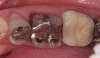
A common form of gastric erosion results from regurgitation. It presents with prominent involvement of the maxillary anterior lingual surfaces (Figure 6). Erosion starts at the gingival margin and reaches to the incisal edge. Gastric reflux disease (or “GERD”) can also cause erosion, mostly on the mandibular posterior teeth. Ditching around existing restorations may also be noted upon examination (Figure 7). A habitual pattern of “Coke swishing” may also produce varied areas of erosion.5
By understanding abrasion, erosion, and abfraction, an astute clinician can begin to recognize that pathologic wear is a multifactorial problem. There will almost always be combinations of occlusal stress and disharmony, erosion, abrasion, parafunction, and attrition (Figure 8). Which components and the influence of each will be unique to every patient, and clinicians should diagnose and treat the cause rather than the symptom. Treating only the symptom will often allow the problem to repeat itself as subsequent dental restorations succumb to the same fate as the natural dentition.
Restorative Implications of the Worn Dentition
Excessive and uncontrolled wear of the dentition often requires extensive restorative dentistry. Management of a complicated restorative situation can often times seem overwhelming. Patients with a worn dentition frequently seek care for esthetic reasons, because the wear often affects their anterior teeth. The author suggests the following considerations in such cases:
Proper form (esthetics) follows proper function. Understanding the parameters that make anterior teeth functional facilitates creating a beautiful smile.
If teeth are in need of restoration due to wear and breakdown, the patient’s current masticatory system is not working properly.
Failure to follow function often leads to failed form or esthetics, particularly in the case of a worn dentition.
If wear has resulted in extensive loss of tooth structure, the teeth have most likely changed position and are now probably in the wrong functional position, eg, active eruption of teeth as wear progresses.15
For instance, the patient in Figure 9 has lost nearly 40% to 50% of his maxillary anterior tooth structure, yet the teeth are still in full intercuspation due to continued eruption as tooth structure is lost due to wear. As wear occurs on the lingual aspects of maxillary anterior teeth, teeth drift to the lingual, which constricts the envelope of function. Wear on the lower incisors can lead to super eruption, which increases the overjet/overbite relationship. Both of these alterations in tooth position steepen the anterior guidance and are less than desirable situations with patients with worn dentitions.
When restoring teeth due to wear, restorations should be placed into a stable functional relationship. Properly positioned and functionally correct anterior teeth are essential to restorative success and occlusal stability. Occlusal requirements that need to be met in restoring a worn dentition are16: (1) even, stable holding contacts on all teeth with both TMJs seated in CR; (2) anterior guidance in harmony with the anterior envelope of function; (3) immediate disclusion of posterior teeth in protrusion; (4) immediate disclusion of posterior teeth in lateral excursive movements.
In the case of a worn dentition, it becomes increasingly critical to restore the teeth meeting these functional requirements and to consider restoring the anterior teeth 3-dimensionally. Specifically, this involves placing the maxillary anterior incisal edges in the proper position. This edge position, both vertically and horizontally, affects the envelope of function, anterior guidance, posterior disclusion, esthetics, phonetics, neutral zone harmony, and lip comfort.
Envelope of Function and Anterior Guidance
The envelope of function is neuromuscularly programmed as the teeth erupt into the neutral zone. During this time, the incisal edge position is established, dictating the anterior guidance. The combination of lingual contour and maxillary anterior inclination influences the relationship between anterior guidance and envelope of function. It is important to note that improper incisal edge position can allow for correct anterior guidance, yet still interfere with the envelope of function.1
Vertical Incisal Edge Position
The restoration of a worn dentition often begins with considering the esthetics of the smile, specifically the appearance of the maxillary central incisors. These teeth influence symmetry and the proportion of the smile and set the functional framework for the envelope of function, anterior guidance, and phonetics.17 Determining the correct tooth length in a patient with a worn dentition can be difficult because known references may have been lost, and the teeth often migrate to an incorrect functional position. Changes can only be estimated and tested during the provisional phase of treatment. When adding length to worn teeth, the dentist should consider adding it both gingivally and incisally. Despite presence of wear, the incisal edge is often at the correct vertical length position. The correct vertical length of a maxillary central incisor should be between 10.5 mm and 11.5 mm.18 The “rest position” of the patient can also help determine the incisal edge position. Vig and Brundo determined that an average of 1.91 mm to 3.4 mm of tooth structure should be visible in the rest position. This amount should decrease with age as gravity pulls the soft tissues toward the ground.19
The “e” position is another useful reference when determining central incisor length. This position is created by the patient pronouncing a long “e” sound, and the relative position of the lips to the incisal edge is evaluated. Ideally, the incisal edge will be between 50% and 70% of the distance between the upper and lower lips.
The ideal length of the maxillary central incisors is approximately 10.5 mm to 11 mm. The rest and “e” positions are then used to determine the approximate vertical position to place the incisal edge. A common restorative error is adding excessive length to the centrals thereby causing envelope of function issues and lip closure path interferences and threatening the balance of the tongue and peri-oral muscles, referred to as the “neutral zone.”20
Horizontal Incisal Edge Position
The horizontal edge position of the incisors may be one of the most critical factors to correctly determine when treating a worn dentition. The horizontal position must be in harmony with the envelope of function and lip closure path, and also be phonetically correct. Lingualization errors will lead to an anterior–posterior constriction in the patient’s envelope of function. This occurs as the maxillary edges interfere with the lower incisors’ arc of closure.21 Additionally, the maxillary horizontal edge position influences production of “f,” “v,” and “s” sounds, and errors in its placement can cause speech difficulty and speech impediments.
The “tip-down” smile and 90º positions are used to evaluate the horizontal position of the maxillary incisal edge. This edge should have a trajectory toward the inner vermillion border (wet/dry line) of the lower lip.
Changes in incisal edge position can only be estimated with a diagnostic wax-up and must be tested with properly contoured provisional restorations. This will verify esthetic, functional, and phonetic success for the patient, and the provisionals will provide a blueprint for the laboratory when creating the final restorations.
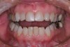
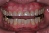
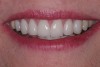
Case Example
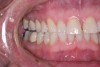
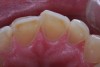
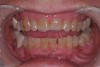
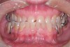
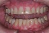
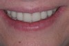
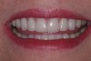
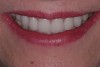
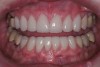
A patient presented with advanced generalized wear of her anterior teeth (Figure 10 and Figure 11). She was displeased with their overall appearance because of their color and wear (Figure 12). A complete examination was performed, revealing instability in her temporomandibular lateral poles bilaterally, sore muscles of mastication, advanced wear, a CR/MIP discrepancy, and loss of her anterior guidance due to the wear. Although the topic is beyond the scope of this article, the patient was also screened for possible sleep apnea. This included an evaluation of the Mallampati score, previous sleep therapy evaluation or treatment, snoring history, an evaluation of her neck size, her weight status, and the presence of the tonsils and their size. In every case, if this clinician suspects airway obstruction to be playing a role in tooth wear issues, the patient is referred to a sleep physician. The patient in this case displayed few apnea risk factors, and the patient’s anterior wear facets fit together like a “lock and key” pattern seen in parafunctional activity. Splint therapy was initiated to stabilize the joints and muscles. A repeatable CR position was verified through load testing. At this point diagnostic models, photographs, a CR bite record, and a facebow were taken and recorded.
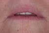
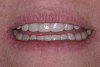
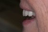
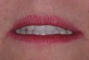
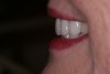
With the degree of wear and instability present, it was critical to place the incisal edges in their correct position for function and esthetics. The patient’s rest position photograph showed nearly the correct amount of tooth display (Figure 13), and the “e” position photograph showed the edge to be at the 60% mark (Figure 14). This meant that very little or about 0.5 mm of tooth length could be added to the incisal edges to gain length. Any additional length would need to come from the gingiva through crown lengthening. The horizontal position was determined to be acceptable based on the tip-down and 90º smile photographs (Figure 15). Figure 16 shows that 1.5 mm of crown lengthening would be necessary to achieve a desirable width/length ratio.
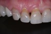
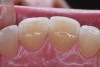
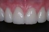
Treatment for this patient included posterior reductive equilibration and additive anterior equilibration (restoration) to eliminate the CR/MIP interferences and to meet the requirements of occlusal stability. Teeth Nos. 5 through 11 and 22 through 27 were prepared for full-coverage lithium-disilicate restorations. Crestal bone heights were evaluated. Soft-tissue crown lengthening was performed on the maxillary anterior teeth by a soft-tissue diode as needed. The esthetic, functional, and phonetic changes were evaluated and confirmed in the provisional restorative phase (Figure 17 through Figure 21).
Once approved, impressions of the provisionals were taken so the laboratory could precisely copy the 3D position of the anterior teeth as successfully proven in the provisionals. The postoperative result and final functional photographs are shown in Figure 22 through Figure 29. Posterior treatment can now be completed in segments as necessary. Posterior morphology will be developed in harmony with the now corrected anterior contour and functional parameters. The fulfillment of the previously mentioned requirements of occlusal stability were evaluated and refined in the final restorations. The patient was placed in a posttreatment dual-arch B splint appliance to help manage any further parafunctional forces should they occur.
Conclusion
The worn dentition provides many challenges for restorative dentists. The cause of the patient’s wear-related condition must be diagnosed. Many failures in restorative care can be linked back to functional issues that were unresolved and undiagnosed. Meeting the requirements of occlusal stability in wear cases will help patients have esthetic, functional, and phonetic success.
Acknowledgment
The author would like to thank his ceramist Mike Felganhauer of DAL Signature Restorations for his artistic efforts in this case.
Disclosure
The author has no relevant financial relationships to disclose.
References
1. Dawson PE. Functional Occlusion: From TMJ to Smile Design. 1st ed. St. Louis, MO: Mosby, Inc.; 2007:1-648.
2. Larson TD. Tooth wear: when to treat, why, and how. Part One. Northwest Dent. 2009;88(5):31-38.
3. Newman MG, Takei HH, Carranza FA. Carranza’s Clinical Periodontology, Volume 1. Philadelphia, PA: WB Saunders Company; 2002:438-439.
4. Niedermeier W. Parameters of tooth mobility in cases of normal function and functional disorders of the masticatory system. J Oral Rehabil. 1993;20 (2):189-202.
5. Abrahamsen TC. The worn dentition—pathognomonic patterns of abrasion and erosion. Int Dent J. 2005;55(4 suppl 1):S268-S276.
6. Shetty S, Pitti V, Satish Babu CL, et al. Bruxism: A literature review. J Indian Prosthodont Soc. 2010;10 (3):141-148.
7. Hess LA. Relevance of occlusion in the golden age of esthetics. Inside Dentistry. 2008;4(2):36-44.
8. Grippo JO, Simring M, Coleman TA. Abfraction, abrasion, biocorrosion, and the enigma of noncarious cervical lesion: a 20-year perspective. J Esthet Restor Dent. 2012;24(1):10-23.
9. Okano N, Baba K, Igarashi Y. Influence of altered occlusal guidance on masticatory muscle during clenching. J Oral Rehabil. 2007;34(9):679-684.
10. Mansour RM, Reynik RJ. In vivo occlusal forces and moments: I. Forces measured in terminal hinge position and associated moments. J Dent Res. 1975;54(1):114-120.
11. Fernandes CP, Chevitarese O. The orientation and direction of rods in dental enamel. J Prosthet Dent. 1991;65(6):793-800.
12. Shetty SM, Shetty RG, Mattigatti S, et al. No carious cervical lesions: abfraction. J Int Oral Health. 2013; 5(5):143-146.
13. Tripathi P, Chopra D, Bagga S. Abfraction Myth or Reality. Journal of Dental and Medical Sciences. 2014;13 (2):70-73.
14. Kanzow P, Wegehaupt FJ, Attin T, Wiegand A. Etiology and pathogenesis of dental erosion. Quintessence Int. 2016;47(4):275-278.
15. Van’t Spijker A, Rodriguez JM, Kreulen CM, et al. Prevalence of tooth wear in adults. Int J Prosthodont. 2009; 22(1):35-42.
16. Hess L. Pre-restorative diagnosis. Inside Dentistry. 2010;6(6):44-54.
17. Hess L. Esthetics conservation: a smile makeover does not always have to be extreme. Inside Dentistry. 2007; 3(2):74-79.
18. Magne P, Gallucci GO, Belser UC. Anatomic crown width/length ratios of unworn and worn maxillary teeth in white subjects. J Prosthet Dent. 2003; 89(5):453-461.
19. Vig RG, Brundo GC. The kinetics of anterior tooth display. J Prosthet Dent. 1978;39(5):502-504.
20. Hess L. Restoring the functional zone: correcting anterior constriction with centric relation based prosthodontics. Inside Dentistry. 2007;3(9):74-79.
21. Cranham J. The horizontal position of the maxillary incisal edge: the key to optimum esthetics, phonetics, and function. Contemporary Esthetics and Restorative Practice. 2006;10(2):22-24.
About the Author
Leonard A. Hess, DDS
Senior Faculty
The Dawson Academy
Private Practice
Monroe, North Carolina