You must be signed in to read the rest of this article.
Registration on CDEWorld is free. You may also login to CDEWorld with your DentalAegis.com account.
Disclosures: The authors had no disclosures to report.
The treatment of congenitally missing lateral incisors generally consists of either canine substitution or prosthetic replacement after idealizing the space. If the choice is made to idealize the space through orthodontics and use some form of prosthetic replacement, there are only a few conservative options, including a resin-bonded bridge or an implant. Of critical importance is choosing the most conservative treatment option available so that there is minimal, if any, modification of the tooth structure.1-4 Once a tooth has been cut, it will require multiple and more aggressive restorative procedures that will necessitate a lifelong commitment from the patient. This cycle should be avoided for as long as possible. Given the need to be conservative, a fixed partial denture is generally a poor option in the adolescent patient due to its inherent aggressiveness in the removal of tooth structure. Therefore, the most conservative options for the replacement of a congenitally missing lateral incisor are utilizing a single tooth implant or some form of resin-bonded bridge.
Single Tooth Implants
Due to the benefit of not having to mechanically prepare the adjacent teeth, single tooth implants have become the most popular treatment option for the replacement of missing anterior teeth. Many studies have demonstrated the long-term success and function of osseointegrated implants.5-9 Although implants may be the ideal option for an adult with congenitally missing lateral incisors, they are not ideal for the growing, adolescent patient. Implants function like ankylosed teeth.10-12 And as a result of continuous growth and development of the alveolar/tooth complex, early placement of osseointegrated implants can lead to infraocclusion, esthetic issues, and periodontal problems.13,14 Therefore, it is imperative to wait until growth cessation has occurred before placing implants. So when is the right time to place dental implants? The most predictable way to monitor facial growth is by evaluating serial cephalometric radiographs taken 6 to 12 months apart.15,16 When the radiographs show no further growth, an implant can be placed. The timing of implant placement after the end of growth generally occurs at about 20 to 21 years of age for men and 16 to 17 years of age for women.16,17 Unfortunately, even if the patient has reached the age range for presumed growth cessation, it does not mean that growth has ceased. Continued growth with eruption and migration of teeth over time has been documented, even into fourth and fifth decade of life, albeit at a much slower rate than the adolescent growth spurt.18-23 This emphasizes the need to wait for the most appropriate time when considering the placement of implants in a young patient. Traditional orthodontic treatment usually begins when a child has lost most of the primary teeth and a majority of the adult teeth have grown in. This generally occurs between the ages of 9 and 14. Given the disparity in time between the completion of orthodontic treatment and when an implant can be placed, some form of interim prosthetic replacement is needed.
Metal Resin-Bonded Bridges as a Permanent Solution
The metal resin-bonded bridge was first popularized by Rochette in 1973.24 He used a cast frame with perforations in combination with resin cement for periodontal splinting. In 1983, at the University of Maryland, Thompson and Livaditis developed the technique of electrolytic etching using Ni-Cr and Co-Cr alloy.25 This technique was found to be almost three times as retentive as the perforated bridge.26 Unfortunately, etching could only be used on non-precious alloys. Noble alloys could not be etched, so instead, electrolytic tin plating was used to facilitate retention. With the advent of the Rocatec system in 1989, which employed the use of a tribochemical silica layer, bond strengths much higher than electroetching or chemical etching were achieved.27,28 Today, a form of tribochemical silica (CoJetTM, 3M ESPE) has been produced that significantly enhances the bonding of resin to metal.29
Even with all of the improvements in bonding metal to resin, metal resin-bonded bridges have exhibited a variable success rate and the highest frequency of failure due to debonding.30-33 According to data from about 25 studies, the mean annual estimated failure rate is somewhere around 4.6% (+/- 1.28%) with a median survival rate of approximately 7.8 years.34,35 Given this lack of long-term clinical success, metal resin-bonded bridges are not to be considered a long term solution for maxillary lateral incisor replacement.
Metal Resin-Bonded Bridges as a Transitional Solution
Out of all of the resin-bonded bridge options, the double-wing metal resin-bonded bridge is ideal for transitioning the patient from adolescence to adulthood when an implant is the final objective. It is very useful as an interim prosthesis due to its ability to be removed to access an edentulous site for the purpose of augmentation and implant placement, its capability to be rebonded until final healing, and its usefulness as a post orthodontic retainer to maintain the ideal root position after orthodontics.36 Another advantage of using a metal alloy resin-bonded bridge is that it is inexpensive and can be made very thin (.7 mm) without decreasing the resistance to flexure.37 All other forms of resin-bonded fixed prostheses require refabrication each time they are removed to access the site for augmentation and implant placement procedures. There are, however, some contraindications when considering resin-bonded bridges. These include deep overbites, proclined teeth, mobility of abutment teeth, and tooth translucency.38 Ideally, a shallow overbite is desirable to allow adequate surface area for bonding and minimize lateral forces on the teeth that can cause debonding.
If a more permanent solution utilizing metal is desired, a “pin-ledge” cantilevered fixed partial denture is an option.39 In order to utilize this form of bridge, pin channels and grooves must be drilled into the lingual surface of the canine tooth, and pins are incorporated into the casting, which is then bonded into place. The advantage of this type of prosthesis, when compared with the double-wing metal resin-bonded bridge, is additional mechanical retention, as well as its ability to remain unaffected by proclination or mobility of the abutments. The chief disadvantage of this type of prosthesis is that it requires tooth preparation, and it will not serve as an orthodontic retainer once the root angulation has been idealized. In addition, drilling the pin hole in the canine tooth negates the objective of tooth structure conservation and will begin the unnecessary life-long commitment to restorative therapy. The final issue is that, if the patient decides to have an implant placed in the future, a provisional will need to be fabricated during the healing phase, after implant placement, if an immediate-load implant is not an option.
Ceramic Resin-Bonded Bridges as a Transitional Solution
In 1991, Kern and colleagues described the use of the all-porcelain resin-bonded bridge, utilizing In-Ceram as the ceramic material of choice.40 Of course, when compared to the use of metal, the big advantage of ceramics was in the esthetics. At that time, In-Ceram was considered a high-strength ceramic (glass-infiltrated alumina), containing 95% aluminum oxide and exhibiting a strength of 400 to 600 MPa.41 Unfortunately, alumina was not amenable to conventional adhesive surface treatment options, such as acid etching. Creating a durable bond required sandblasting to improve wetting of the surface and increase surface area and the use of a composite resin modified with a phosphate monomer (Panavia 21), which bonds to metallic oxides.42 In 2005, Kern evaluated the survival of double-wing versus single-wing In-Ceram resin-bonded bridges and found that the 5-year survival rate was 73.9% for the double-wing group and 92.3% for the single-wing group.43 In the double-wing group, failure was mostly due to fracture of the connector.43 In 2011, Kern and another researcher did a follow-up study on these same bridges and found that the 10-year survival rate of the double-wing group was the same as at 5 years, while the single-wing group had a survival rate of 94.4%.44 Interestingly, when a retainer broke among the double-wing group, the bridge still survived with one wing. More recently, zirconia has been used as a framework for resin-bonded bridges due to its increased flexural strength (somewhere between 900 to 1,200 MPa). In another study, Sasse and colleagues evaluated the 3-year survival of single-wing zirconia resin-bonded bridges bonded with Panavia 2.0. They found a 100% survival rate; however, 2 out of the 30 resin-bonded bridges had debonded, and both were rebonded with no other issues.45 A follow up study at 6 years also demonstrated a survival rate of 100%, with the same two debonded bridges needing to be recemented and one carious lesion needing repair.46 Given zirconia’s strength and effectiveness, it may be a good long-term strategy for a situation in which a patient either does not want an implant or has no room for an implant and does not want to address the issue with orthodontics. With the introduction of transformation-toughened zirconia, many different bonding strategies were developed to help retain the non-etchable surface of zirconia.47-49 It seems that the combination of primers (MDP) and air-abrasion methods tend to produce good long-term bond strength.50-53 In theory, phosphate monomers form chemical bonds with the zirconia, alumina, and metal oxide surfaces and have resin terminal end groups, which enable cohesive bonding to appropriate resin cements.54,55 More recently, glazing techniques on zirconia (ie, the application of acid-etchable glasses/porcelain on zirconia) followed by acid etching with hydrofluoric acid has shown improved bond strengths. Everson, et al. found this increased bond strengths to zirconia, and Valentino et al. found that treatment of zirconia with a glaze of lower fusing porcelain significantly increased the bond strength of a dual-cured resin luting agent to zirconia when compared to conventional airborne particle abrasion with 50 to 110 pm aluminum oxide.56,57 Kitayama et al. found that the internal coating of zirconia dental restorations with silica-based ceramic followed by salinization may be indicated in order to achieve better bonding for clinical success, although obtaining the space for placement of the feldspathic layer is the key to the fit of the prosthesis.58
Case Presentation 1: Patient Wants Implants as a Final Prosthetic Solution
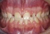
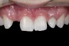
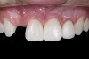
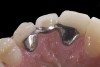
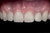
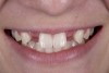
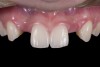
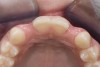
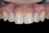
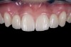
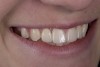
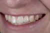
A 14-year-old patient presented to the office with his mother (Figure 1). His chief complaint involved the large spaces between the teeth created by his missing right maxillary lateral incisor and his small left maxillary lateral incisor. He desired to replace the missing tooth with an implant and create a beautiful smile. Upon examination, he was found to have a class I canine and molar relationship, but because he had a tooth size/arch size discrepancy and space distal to the right central incisor, the remaining incisors had drifted to the right. The left maxillary lateral incisor was peg-shaped and in a cross bite position. Studies have shown a clear association between congenitally missing teeth and reduced tooth size.59-62 Because he was only 14-years-old at the time and could not have implants placed until the cessation of growth (somewhere in the vicinity of 22 years old), he was sent to the orthodontist for alignment of the teeth.16,17 After 2 years of orthodontics, the appliances were removed, and his tooth coloration was improved using carbamide peroxide bleaching (Figure 2). Because some form of provisional needed to be placed until he was finished growing, a double-wing metal resin-bonded bridge was chosen. As discussed earlier, this is the ideal transitional prosthesis for patients that have congenitally missing maxillary lateral incisors. The benefits of this type of prosthesis include its ability to be removed and rebonded during the surgical phase of treatment and its ability to retain the roots in their proper position after orthodontic treatment.16 The final plan for the patient was to increase the width of the central and the maxillary left lateral incisor, utilizing porcelain laminate veneers to achieve the appropriate width/length ratio of 80%. A wax-up was created to idealize tooth size, a putty matrix was made from the wax-up to facilitate bonding of the incisors, and a non-precious, double-wing metal resin-bonded bridge was fabricated for lateral incisor replacement. Once the teeth were bonded to ideal size, the “Maryland Bridge” was fabricated from a polyvinyl arch impression with the newly bonded teeth (Figure 3). The metal frame was cast from a non-precious alloy to allow for fabrication of a very thin frame and to create a better surface for bonding. After sandblasting the internal aspect of the frame with CoJet™ silica (3M ESPE), accomplishing salinization, and executing cementation with a dual-cure resin cement, a fairly good adhesion to the frame was anticipated.29 The enamel surface was etched with phosphoric acid for 30 seconds, the primer (Single Bond Plus, 3M ESPE) was applied to both the internal surface of the sandblasted framework and the etched enamel, and the bridge was cemented with RelyX™ ARC (3M Espe) dual-cured resin cement (Figure 4 and Figure 5).
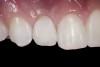
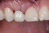
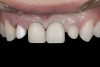
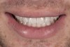
Six years later, the patient was in his fourth year of college when he finally returned for implant placement (Figure 6). Interestingly, the provisional bridge had not come loose since it was bonded, even though the patient had a nail biting habit and had fractured most of the resin-based composite on the central incisors. He had also failed to wear his retainer, and as a result, the upper left lateral incisor had rotated slightly (Figure 6). In the right lateral incisor site, there was a horizontal deficiency in the bone and overlying soft tissue. A cone-beam computed tomography (CBCT) image was taken, and the area was evaluated for implant placement. There was enough native bone to place a 3 mm implant, but a connective tissue graft was needed to rebuild the deficient ridge. Unfortunately, when the patient presented for implant placement, it was during a holiday break from school and he needed to study. Therefore, the patient did not want to simultaneously augment with a connective tissue graft at the time of implant placement due to the pain associated with treatment. A 3.0 x 10 mm Astra Tech implant was placed, and the resin-bonded bridge was rebonded, utilizing the CoJet Silica, salinization, and the same dual-cured resin cement. Four months later, the patient returned for the connective tissue graft (Figure 7). The bridge was removed, the graft was performed utilizing a tunnel technique, and the bridge was rebonded as described earlier (Figure 8). After another four months, the patient returned during a two-week break. At this time, the implant was exposed, the tissue was manipulated with provisionals, and the final restorative work was completed (Figure 10 and Figure 11).
Case Presentation 2: Patient Does Not Want to Have an Implant Placed
Some patients do not want an implant due to the need for surgical intervention, and others may not have enough space for implant placement. A 15-year-old patient presented to the office after completion of orthodontic treatment to idealize the spacing of her teeth and improve her occlusal relationship (Figure 12 through Figure 14). Because both of her maxillary lateral incisors were missing, the patient was wearing a retainer with lateral incisor pontics. She had a busy school schedule, but expressed her desire for a fixed restorative option. All of her options were discussed, and the patient was informed that if she wanted an implant, she would have to wait for at least 3 to 4 years for completion of growth. The patient was not sure if or when she wanted to have an implant placed in the future, especially considering her busy schedule and desire to attend college after high school. Considering her age and the need to be conservative, a single-wing zirconia Maryland Bridge was chosen as the ideal prosthetic replacement option. Because bonding a non-etchable and smooth surface such as zirconia requires chemical adhesion, it was decided to use a modified technique to make the bridge more retentive. One of the ways to improve adhesion of a zirconia bridge is to use an etchable feldspathic ceramic layer on the internal surface of the zirconia retainer.56-58 Unfortunately, it can be difficult to determine the thickness of the ceramic and ensure accurate seating of the restoration.
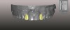
An approach utilizing CAD/CAM was employed to fabricate an accurate fitting, retentive, and esthetic ceramic single-wing resin-bonded bridge. Impressions were taken of the patient and scanned in the laboratory using maximum intensity projection. The scanned models were then used to design the final prosthetic frames utilizing EXOCAD software. The retainer positions and extensions were designed on the models based on the occlusal clearance (Figure 15). Next, the replacement tooth forms were chosen from a virtual library (Figure 16), and try-in PMMA bridge prototypes were milled in Primotec USA PMMA. This allowed for try-in of the design, adjustment of the contacts, and intraoral evaluation of the ridge contact (Figure 17). Once tried and idealized for both fit and occlusion, the design was sent back to the lab to rescan. Utilizing the software, a cut back was made on the lateral incisor (Figure 18), and a 0.3mm space was also virtually designed on the internal surface of the retainer wing to allow layering with feldspathic ceramic (Figure 19). The 0.2 mm internal edge of the wing was designed to be left intact to allow for precise seating and verification of fit on the model. After the frame was milled in zirconia (Zirconzahn, Prettau), it was verified on the model, and high fusing margin porcelain (Noritake CZR) was mixed with a clear utility wax and applied to the internal 0.3 mm depression. This was baked in the oven at a high temperature, allowing the wax to burn out and leaving the feldspathic ceramic fused to the internal surface of the retainer. Once cooled, this was checked for precise fitting on the model and adjustments were made to ensure full seating.
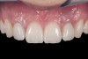
When the patient returned, the fit was evaluated in the mouth. Once verified, the internal surface of the framework was etched with a 9.5% hydrofluoric acid for 90 seconds and then salinized. After etching the enamel surface with phosphoric acid for 30 seconds and applying the primer (Single Bond Plus, 3M ESPE) to both the internal surface of the framework and the enamel, the zirconia bridge was bonded with a dual-cure resin cement (RelyX™ ARC, 3M ESPE). After the procedure, the patient ended up with a long-term, high-strength esthetic restoration advantaged by the bonding potential of fledspathic ceramic (Figure 20 through Figure 24). Six years after placement, the prosthesis had remained in place with no complications.
Discussion
This article examined the options and considerations for adolescent patients with congenitally missing lateral incisors and presented a modified technique for enhanced adhesion. When treating the adolescent patient and deciding what type of resin-bonded bridge to use, one must consider the most conservative approach as well as the objectives of future treatment. Both metal and ceramic resin-bonded bridges perform well, but in different capacities. Ultimately, the final plan will be the primary driver in deciding what type of bridge to use.
Acknowledgment
I would like to thank Emanuele Piazzera, MDT, for his amazing laboratory skills and his vast knowledge of CAD/CAM technology.
References
1. Kavadia S, Papadiochou S, Papadiochos I, Zafiriadis L. Agenesis of maxillary lateral incisors: A global overview of the clinical problem. Orthodontics (Chic). 2011; 12(4):296–317.
2. Kokich VO, Kinzer GA, Janakievski J. Congenitally missing maxillary lateral incisors: restorative replacement. Am J Orthod Dentofacial Orthop. 2011;139(4):435-439.
3. Avila ED, Molon RS, Assis Mollo F, et al. Multidisciplinary approach for the aesthetic treatment of maxillary lateral incisors agenesis: thinking about implants? Oral Surg Oral Med Oral Pathol Oral Radiol. 2012;114 (5):e22–e28.
4. Jackson BJ, Slavin MR. Treatment of congenitally missing maxillary lateral incisors: an interdisciplinary approach. J Oral Implantol. 2013;39(2):187-192.
5. Belser UC, Schmid B, Higginbottom F, Buser D. Outcome analysis of implant restorations located in the anterior maxilla: a review of the recent literature. Int J Oral Maxillofac Implants. 2004;19(Suppl):30–42.
6. Zarone F, Sorrentino R, Vaccaro F, Russo S. Prosthetic treatment of maxillary lateral incisor agenesis with osseointegrated implants: a 24-39-month prospective clinical study. Clin Oral Implants Res. 2006;17(1):94–101.
7. Henry PJ, Laney WR, Jemt T, et al. Osseointegrated implants for single-tooth replacement: a prospective 5-year multicenter study. Int J Oral Maxillofac Implants. 1996;11(4):450–455.
8. Reddy MS, O’Neal SJ, Haigh S, Aponte-Wesson R, Geurs NC. Initial clinical efficacy of 3-mm implants immediately placed into function in conditions of limited spacing. Int J Oral Maxillofac Implants. 2008;23 (2):281–288.
9. Romeo E, Lops D, Amorfini L, Chiapasco M, Ghisolfi M, Vogel G. Clinical and radiographic evaluation of small-diameter (3.3-mm) implants followed for 1-7 years: a longitudinal study. Clin Oral Implants Res. 2006;17(2):139–148.
10. Thilander B, Odman J, Grondahl K, Lekholm U. Aspects on osseointegrated implants in growing jaws. A biometric and radiographic study in the young pig. Eur J Orthod. 1992;14(2):99-109.
11. Odman J, Grondahl K, Lekholm U, Thilander B. The effect of osseointegrated implants on the dento-alveolar development. A clinical and radiographic study in growing pigs. Eur J Orthod. 1991;13(4):279-286.
12. Wehrbein H, Diedrich P. Endosseous titanium implants during and after orthodontic load–an experimental study in the dog. Clin Oral Implants Res. 1993;4(2):76-82.
13. Brugnolo E, Mazzocco C, Cordioll G, Majzoub Z. Clinical and radiographic findings following placement of single-tooth implants in young patients – case reports. Int J Periodontcs Rest Dent. 1996;16(5);421-433.
14. Thilander B, Odman J, Grondahl K, Friberg B. Osseointegrated implants in adolscents. An alternative in replacing missing teeth? Eur J Orthod. 1994;16 (2):84-95.
15. Kokich VG. Managing orthodontic-restorative treatment for the adolescent patient. In: McNamara JA, Brudon WI, eds. Orthodontics and Dentofacail Orthopedics. Ann Arbor, Mich: Needham Press; 2001:P423-P452.
16. Fudalej P, Kokich BG, Leroux B. Determining the cessation of vertical growth of the craniofacial structures to facilitate placement of single-tooth implants. Am J Orthod Dentofacial Orthop. 2007;131(4 Suppl):S59-S67.
17. Kokich Jr. VO, Kinzer GA, Janikievski J. Congenitally missing maxillary lateral incisors: restorative replacement. Counterpoint. Am J Orthod Dentofacial Orthop. 2011;139(4):435-445.
18. Forseberg CM, Eliasson S, Westergren H. Face height and tooth eruption in adults–a 20-year follow-up investigation. Eur J Orthod. 1991;13(4):249-254.
19. Behrents RG. A treatise of continuum of growth in the aging craniofacial skeleton (thesis). Ann Arbor, Michigan: University of Michigan; 1984.
20. Sarnas KV, Solow B. Early adult changes in the skeletal and soft-tissue profile. Eur J Orthod. 1980; 2(1):1-12.
21. Bishara SE, Treder JE, Jakobsen JR. Facial and dental changes in adulthood. Am J Orthod Dentofacial Orthop. 1994;106(2);175-186.
22. Bondevik O. Growth changes in the cranial base and the face: A longitudinal cephalometric study of linear and angular changes in adult Norwegians. Eur J Orthod. 1995;17(6):525-532.
23. Daftary F, Mahallati R, Bahat O, Sullivan RM. Lifelong craniofacial growth and the implications for osseointegrated implants. Int J Oral Maxillofac Implants. 2013;28(1):163-169.
24. Rochette AL. Attachment of a splint to enamel of lower anterior teeth. J Prosthet Dent. 1973;30(4 Pt 1):418-423.
25. Livaditis GJ, Thompson VP. Etched castings: an improved retentive mechanism for resin-bonded retainers. J Prosthet Dent. 1982;47(1):52-58.
26. Dhillon M, Fenton AH, Watson PE. Bond strength of composite to perforated and etch metal surfaces. J Dent Rest. 1983;62:304.
27. Caeg C, Leinfelder KF, Lacefield WR, Bell W. Effectiveness of a method used in bonding resins to metal. J Prosthet Dent. 1990;64(1):37-41.
28. Ishijima T, Caputo AA, Mito R. Adhesion of resin to casting alloys. J Prothet Dent. 1992;67(4):445-449.
29. Matinlinna JP, Vallittu PK. Silane based concepts on bonding resin composite to metal. J Contemp Dent Pract. 2007;8(2):1-8.
30. Williams VD, Thayer KE, Denehy GE, Boyer DB. Cast metal, resin-bonded prostheses: a 10-year retrospective study. J Prosthet Dent. 1989;62(1):436-441.
31. Hansson O. Clinical results with resin-bonded prostheses and an adhesive cement. Quintessence Int. 1994;25(2):125-132.
32. Priest GF. Failure rates of restorations for single-tooth replacement. Int J Prosthodont. 1996;9(1):38-45.
33. Probster B, Henrich GM. 11-year follow-up study of resin-bonded fixed partial dentures. Int J Prosthodont. 1997;10(3):259-268.
34. Miettinen M, Millar BJ. A review of the success and failure characteristics of resin-bonded bridges. Br Dent J. 2013;215(2):E3.
35. Dunne SM, Millar BJ. A longitudinal study of the clinical performance of resin bonded bridges and splints. Br Dent J. 1993;174(11):404-411.
36. Olsen TM, Kokich VG Sr. Postorthodontic root approximation after opening space for maxillary lateral incisor implants. Am J Orthod Dentofacial Orthop. 2010;137(2):158-159.
37. Ibrahim AA, Byrne D, Hussey DI, Claffey N. Bond strengths of maxillary anterior base metal resin-bonded retainers with different thicknesses. J Prosthet Dent. 1997;78(3):281-285.
38. Creugers NH, Kayser AF, Van’t Hof MA. A seven-and-a-half-year survival study of resin-bonded bridges. J Dent Res. 1992;71(11):1822-1825.
39. Small BW. The use of cast gold pinledge retainers with pontics as an esthetic and functional restorative option in the maxillary anterior. Gen Dent. 2004;52 (1):18-20.
40. Kern M, Knode H, Strubb JR. The all-porcelain, resin-bonded bridge. Quintessance Int. 1991;22(4):257-262.
41. Ironside, JG, Swain MV. Ceramics in Dental Restorations – A Review and Critical Issues. Journal of the Australasian Ceramic Society. 1998;34(2):78-91.
42. Kern M, Thompson VP. Bonding to glass infiltrated alumina ceramic: adhesive methods and their durability. J Prosthet Dent. 1995;73(3):240-249.
43. Kern, M. Clinical long-term survival of two-retainer and single-retainer all-ceramic resin-bonded fixed partial dentures. Quintessence Int. 2005;36(2):141-147.
44. Kern M, Sasse M. Ten-year survival of anterior all ceramic resin-bonded fixed dental prostheses. J Adhes Dent. 2011;13(5):407-410.
45. Sasse M, Eschback S, Kern M. Randomized clinical trial on single retainer all-ceramic resin-bonded fixed partial dentures: influence of the bonding system after up to 55 months. J Dent. 2012;40(9):783-786.
46. Sasse M, Kern M. Survival of anterior cantilevered all-ceramic resin-bonded fixed dental prostheses made from zirconia ceramic. J Dent. 2014;42(6):660-663.
47. Blaltz MB, Sadan A, Kern, M. Resin-ceramic bonding: a review of the literature. J Prosthet Dent. 2003; 89(3):268-274.
48. Della Bona A, Borba M, Benetti P, Cecchetti D. Effect of surface treatments on the bond strength of a zirconia-reinforced ceramic to composite resin. Braz Oral Res. 2007;21(1):10-15.
49. Donassollo TA, Demarco FF, Della Bona A. Resin bond strength to a zirconia-reinforced ceramic after different surface treatments. Gen Dent. 2007;57(4):374-379.
50. Yang B, Barloi A, Kern M. Influence of air-abrasion on zirconia ceramic bonding using an adhesive composite resin. Dent Mater. 2010;26(1):44–50.
51. Lehmann F, Kern M. Durability of resin bonding to zirconia ceramic using different primers. J Adhes Dent. 2009;11(6):479-483.
52. Yun JY, Ha SR, Lee JB, Kim SH. Effect of sandblasting and various metal primers on the shear bond strength of resin cement to Y-TZP ceramic. Dent Mater. 2010;26(7):650-658.
53. Silva LH, Costa AK, Queiroz JR, Bottino MA, Valandro LF. Ceramic primer heat-treatment effect on resin cement/Y-TZP bond strength. Oper Dent. 2012;37 (6):634–640.
54. Aboushelib MN, Matinlinna JP, Salameh Z, Ounsi H. Innovations in bonding to zirconia-based materials: Part I. Dent Mater. 2008;24(9):1268–1272.
55. Yoshida K, Tsuo Y, Atsuta M. Bonding of dual-cured resin cement to zirconia ceramic using phosphate acid ester monomer and zirconate coupler. J Biomed Mater Res B Appl Biomater. 2006;77(1):28–33.
56. Everson P, Addison O, Palin WM, Burke FJ. Improved bonding of zirconia susbstructures to resin using a “glaze-on” technique. J Dent. 2012;40(44):347-351.
57. Valentino TA, Borges GA, Borges LH, Platt JA, Correr-Sobrinho L. Influence of glazed zirconia on dual-cure luting agent bond strength. Oper Dent. 2012;37 (2):181-187.
58. Kitayama S, Nikaido T, Ikeda M, Alireza S, miura H, Tagami J. Internal coating of zirconia restoration with silica-based ceramic improves bonding of resin cement to dental zirconia ceramic. Biomed Mater Eng. 2010; 20(2):77-87.
59. Lyngstadaas SP, Nordbo H, Gedde-Dahl T Jr, Thrane PS. On the genetics of hypodontia and microdontia: synergism or allelism of major genes in a family with six affected members. J Med Genet. 1996;33(2):137–142.
60. Wu CC, Wong RW, Hägg EU. A review of hypodontia: The possible etiologies and orthodontic, surgical and restorative treatment options: conventional and futuristic. Hong Kong Dent J. 2007;4(2):113–121.
61. Brook AH. A unifying aetiological explanation for anomalies of human tooth number and size. Arch Oral Biol. 1984;29(5):373–378.
62. Gungor AY, Turkkahraman H. Tooth sizes in nonsyndromic hypodontia patients. Angle Orthod. 2013; 83(1):16–21.
About the Author
TAL MORR, DMD, MSD
Owner
TM Prosthodontics
Miami, Florida