You must be signed in to read the rest of this article.
Registration on CDEWorld is free. You may also login to CDEWorld with your DentalAegis.com account.
Whether good or bad, people in modern society continue to seek solutions for esthetic- and aging-related problems. This author has often been told by patients that they refuse to suffer a dental fate similar to that experienced by their parents. What is often described by the patient as being only a “cosmetic or esthetic” complaint is often complicated. A concern over the look of worn, chipped, or cracked teeth is often a complex problem rooted in functionally derived issues. Failure by the dentist to recognize a functional problem and simply treat an esthetic issue can certainly lead to failure. This issue can often be compounded by the patient’s limited focus on only the visible “smiling” (anterior) teeth, and this can be further fueled by misinformation through creative dental marketing, often forcing their provider into difficult situations. Many times, these straightforward problems for the patient present significant treatment planning difficulties for the dentist. For example, a patient wanting longer teeth for a more youthful look often does not want to be burdened with complex choices or long-term treatment plans. However, it is at these times that the patient-management skills of the dentist are paramount, as adding 2 mm of incisal length to the maxillary anterior teeth can be the beginning of esthetic, functional, and phonetic errors. Unhappy and dissatisfied patients are not only a problem for those directly involved in their care; when trust in the profession is lost by any single patient, the loss is carried by all.
Planning for Functional Success
Managing complicated restorative situations can often be overwhelming. When such a situation arises, it is often helpful to break the problem down into manageable components. Predictable success is usually the product of a consistent methodology in the diagnosis and planning stages. It also allows both the patient and dentist participation in the exploratory process, and will aid in both parties’ commitment to the outcome.
The data collected before active restoration should include a complete medical and dental history, preoperative photographs, necessary radiographs, periodontal examination, diagnostic impressions, face-bow record, bite registration in centric relation and maximum intercuspation, and a complete maxillofacial exam with emphasis on the condition of the temporomandibular joint.1 Although the topic is beyond the scope of this article, it is important to reinforce the restorative philosophy of this author with regard to centric relation. When cases involve anterior reconstruction, the need has often arisen because of some form of instability in the patient’s occlusion. Therefore, effort is put forth to determine the health of the joints and to begin the restorative phase with the joints in centric relation. If stability is to be regained through the proposed treatment, it must be evident in the joint to allow long-term success.
Special emphasis must be placed on pretreatment photographs obtained in the planning stages. This author employs approximately 22 different photographic shots during this phase. With regards to the incisal edge position, seven photographic shots will be highlighted (Figure 1 through Figure 7).
Factors Influenced By and Influencing the Position of the Incisal Edges
Anterior Envelope of Function and Anterior Guidance
The envelope of function determines the incisal edge position, which then dictates the anterior guidance. The combination of the lingual contours, position, and inclination of the maxillary anterior teeth influence whether the relationship of the anterior guidance and envelope of function is harmonious. Improper positioning of the incisal edges can allow proper anterior guidance, yet still interfere with the envelope of function.2
Neutral Zone
The neutral zone is the perioral complex composed of the soft tissues and muscles surrounding the mouth, and the opposite pressure exerted by the tongue.3 Prosthodontic errors that violate the neutral zone can be very uncomfortable for a patient and start a chain reaction of compensations. Lip-closure paths and muscle-induced function against the teeth must be considered.
Phonetics
The interaction of the lower lip against the incisal edges is critical for many proper speech sounds. Misplacement of the incisal edge can create speech difficulties and minor impediments.
Determining the Length of the Incisors (Vertical Incisal Edge Position)
The maxillary incisors are often the starting point for smile rehabilitation. Diagnostic planning of this aspect should be precise because it influences symmetry, proportion, and functional parameters of the adjacent anterior teeth.4 The final, restored length will be influenced by a variety of individual factors. These would include the lower lip position during smiling, the lip positions at rest, the lip positions in repose, upper lip characteristics, soft tissue characteristics, the envelope of function, and facial proportion.5 Depending upon the preparation style or restorative product used, the length will set the foundation for the width-to-length ratio (an accepted ratio for width-to-length is approximately 80%).6,7 This arrangement also lays the foundation for establishment of the anterior “golden proportion.” Ideally, the width of the lateral incisor would be assigned a value of 1, the canine a value of 0.6, and the central incisor a value of 1.6. This is an apparent measurement and should only be relied on when viewing from the direct anterior. The canine tooth’s value of 0.6 from the anterior is dictated from the facial line angles and height of contour. When viewed laterally, the canine would have a value much greater than 0.6.8
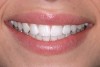
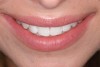
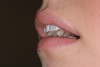
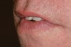
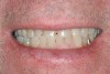
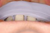
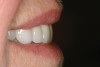
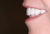
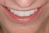
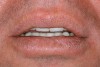
Determining the correct length can be very difficult in patients who present with worn dentition. The length can only be estimated from known references and the collected data. The result must then be tested by the patient during the provisional phase. Studies of crown lengths have shown that unworn central incisors average 11.69 mm in length, and worn centrals average 10.67 mm in length.9 Figure 4 illustrates a patient in the rest position. This position is accomplished by having a patient close their lips with relaxed facial muscles, and then slightly open their lips as if to breathe through their mouth. In this position, Vig and Brundo10 determined that men show an average of 1.91 mm of central incisor and women show an average of 3.4 mm of central incisor. These amounts will decrease with advancing age because the soft tissues are more affected by gravity.10 Figure 6 represents a patient in the “E” position. This is a repose position created by the lips and musculature when having the patient say “E.” It is similar to a smile position, but simulates the position of the soft tissue in a more functioning position. Ideally, the incisal edge of the central incisor will fall 50% to 60% of the distance between the upper and lower lip.
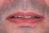
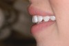
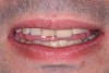
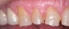
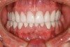
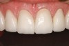
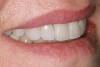
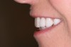
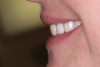
This author has found that a good starting point for the central incisor length is 10.5 mm. The “E” and rest positions are then used to determine the approximate vertical position to place the incisal edge. In many cases involving tooth wear, the patient will want longer-looking teeth. Simply adding length to the incisal to achieve 10.5 mm will often result in encroachment on the envelope of function and the neutral zone. Phonetically, the position of the incisal edge is important when considering the lower lip. The lower lip must interact with the incisal edge in a natural, effortless fashion. This is evident during the pronunciation of words beginning in the letters “V” and “F” (Figure 8). The incisal edge contact should occur in the moist or inner vermilion border of the lip and not the cutaneous or dry portion. This error can often been seen in restorations that are too long incisally.6 The patient in Figure 9 has obviously been occlusally compromised and has extreme wear. However, consider the patient’s current incisal edge location seen in the “E” position (Figure 10). The current position is very close to the ideal vertical position. If an ideal incisor length is to be achieved, the length must be captured from the apical direction. This particular case is an example of passive delayed eruption. As the teeth slowly wear, the alveolar process migrates to keep pace with the tooth loss. The cementoenamel junction, soft tissue attachments, and incisal edge position stay anatomically in place as the process migrates.11 The final restorative process for this patient included hard tissue crown lengthening to recapture added tooth length (Figure 11) and maintenance of the current incisal edge position. The proper maxillary occlusal plane, envelope of function, and anterior guidance were then restored. All of the above parameters were established in the diagnostic phase and then tested in long-term provisionals. The final restorative result can be seen in Figure 12 through Figure 14.
Determining the Horizontal Position of the Incisal Edge
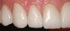
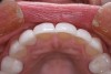
Establishing the proper horizontal incisal edge position may be one of the most important, yet most commonly overlooked, factors in anterior reconstruction. This position must accommodate the patient’s envelope of function and neutral zone. A position too far to the facial can lead to interference as the lower lip closes to seal against the upper lip. A facialization error can also affect the function of the upper lip, causing the lip to have to “work around” the incisal edge. Many times the patient will complain of muscle fatigue and a “not quite right” feeling during function. A lingualization error of the incisal edge can lead to an anterio-posterior constriction in the patient’s envelope of function. This would occur as the edges interfere with the lower incisors’ arc of closure.3,12 Similarity exists with the vertical incisal edge position with regard to phonetic issues. A horizontal discrepancy can interfere with the production of “F” and “V” sounds. In addition, the horizontal edge position must accommodate production of the “S” sound. These sounds are created by the squeezing of air between the upper and lower incisors. Difficulty in creating this sound can be very frustrating for the patient.2,13 This author has found it to be more common to err with the edge position to the facial. This is usually caused by the preparation design error of under-reduction and failure to reduce anterior teeth in three planes. When the middle and incisal thirds are under-reduced, the laboratory technician has no choice but to fabricate an over-contoured restoration. Reduction stents can be produced from the diagnostic wax-up to aid in proper reduction depths (Figure 15). The final restorations should exhibit a three-plane convex contour as seen in natural teeth (Figure 16 and Figure 17).
Communication with the Dental Ceramist
To create predictable success, the laboratory and ceramist must also be a vital part of the process. The technician needs to have a working mastery of functional smile design. Only then can the dentist and technician communicate effectively. A functional understanding of occlusion and smile design cannot stop once a case leaves the dental practice. Therefore, it is the responsibility of the dentist to ensure the dissemination of information to the ceramist.
The necessary information would include preoperative and preparation photographs, and photographs of the provisional restorations. Impressions of the provisional restorations are a necessity. The ceramist must have a reference point to begin the functional contours of the final restorations. Putty stents can be made of the provisionals to duplicate exactly the position of the incisal edges and functional contours of the teeth. In addition, a face-bow of the provisional restorations should be included. The face-bow allows a three-dimensional transfer of the maxilla (this includes the incisal edges) and temporomandibular joint to be transferred to the articulator. The face-bow also allows transfer of the condylar axis of closure to be replicated; this is absolutely necessary when using an open bite record to capture centric relation.2 The alternative is simply guesswork by the laboratory, which typically results in heavy adjustment of the restorations.
Detecting Errors in Incisal Position
It is important to have a command of this knowledge not only to plan a case, but also to aid in recognizing mistakes.
Facial Positional Error
When the incisal edge is restored too far to the facial, the patient may complain of teeth that feel too long, dry, or in the way of the upper lip. The lower lip may feel overworked or that it must over-extend forward to meet the incisal edge while pronouncing “F” or “V.” Evidence may be found that the edges are interacting with the dry or cutaneous portion of the lower lip. In addition, the patient may have trouble producing “S” sounds as the mandible strains to get close to the malpositioned incisal edges. In short, this author has found that a facial error produces mostly complaints in the patient’s feel and phonetic function.
The patient seen in Figure 18 was unable to get comfortable with the restored incisal edge position. The re-treated position seen in Figure 19 through Figure 21 was a subtle, yet profound, change. Because of her thin lip structure (highly muscular), even a small error was detectable and interfered with her lip function.
Lingual Positional Error
Errors of this variety will often be evident from problems associated with restoration failure. It is with this mistake that the edges will hit first in the patient’s arc of closure. Chipped porcelain and debonded restorations may be commonly seen. Problems with provisional restorations breaking or coming off can be an early warning sign that contour changes are needed. Issues with provisionals should not be viewed as an aggravation, but as a chance to refine positions and correct problems before they become set in porcelain. Be cautious when patients complain of feeling that their teeth are hitting wrong even when it only occasionally happens. The patient’s lower lip may also be overworking to the lingual to interact with the lingualized edges. Difficulty may also be encountered during pronunciation of “S” sounds as teeth nip each other in this malposition.
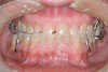
Properly contoured and patient-tested provisional restorations are the only way to test the correctness of the incisal edge position (Figure 22 and Figure 23). Patients often try to rush the process and want to limit the time in provisionals. Proper patient education and production of high-quality temporaries will ease this process and provide the necessary information.
Conclusion
Having a through knowledge and a systematic approach to functional design will lead to predictable restorations. Understanding the requirement for proper incisal edge position will set the foundation for anterior occlusal rehabilitation. By breaking down complex problems into manageable components, treatment planning can become simple and predictable.
Acknowledgment
Dr. Hess would like to thank Dental Arts Signature Laboratory (Peoria, IL) for the fabrication of the restorations seen in this article.
References
1. Hess L. Restoring the functional zone: correcting anterior constriction with centric relation-based prosthodontics. Inside Dentistry. 2007;3(9):74-79.
2. Dawson P. Functional Occlusion: From TMJ to Smile Design. St. Louis, Mo: Mosby; 2006: 141-147, 181.
3. Dawson P. Evaluation, Diagnosis, and Treatment of Occlusal Problems. 2nd Ed. St. Louis, Mo: Mosby; 1989:330.
4. Hess L. Esthetic conservation: a smile makeover does not always have to be extreme. Inside Dentistry. 2007;3(2):74.
5. Hess L. Interdisciplinary synergy: managing complex treatment objectives for a predictable esthetic result. Advanced Esthetics and Interdisciplinary Dentistry. 2006:2(2): 10-18.
6. Chiche GJ, Pinault A. Esthetics of Fixed Anterior Prosthodontics. Hanover Park, IL: Quintessence Publishing Co; 1994:21, 23.
7. Wolfart S, Thormann H, Freitag S, et al. Assessment of dental appearance following changes in incisor proportions. Eur J Oral Sci. 2005;113(2):159-165.
8. Ascheim KW, Dale BG. Esthetic Dentistry: A Clinical Approach to Techniques and Materials. 2nd Ed. St. Louis, Mo.: Mosby; 2000:31.
9. Mange P, Gallucci GO, Belser UC. Anatomic crown width/length ratios of unworn and worn maxillary teeth in white subjects. J Prosthet Dent. 2003:89(5):453-461.
10. Vig RG, Brundo CG. The kinetics of anterior tooth display. J Pros Dent. 1978;39(5): 502-504.
11. Chu S. “Short-tooth syndrome”: diagnosis, etiology, management, and treatment. Calif Dent Assoc J. 2004;32(2):143.
12. Cranham J. The horizontal position of the maxillary incisal edge: the key to optimum esthetics, phonetics and function. Contemporary Esthetics. 2006;10(2):22-24.
13. Hess L. The relevance of occlusion in the Golden Age of esthetics. Inside Dentistry. 2008;4(2):36-44.
About the Author
Leonard A. Hess, DDS
Private Practice
Monroe, North Carolina